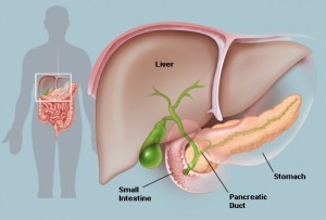
 A research team lead by Professor Ulf Ahlgren and his colleagues is at the forefront of the development of optical projection tomography. Based in Umeå University in Sweden, the team has used this emerging technology to describe embryonic development of the pancreas and the distribution of the islets of Langerhans in the adult pancreas. The team’s findings will aid future research that utilizes modeling of human organs in treating diabetes.
The researchers presented new methods of conducting Optic Projection Tomography in the journal “IEEE Transactions on Medical Imaging.” The team has utilized these methods to perform research into the structure of the adult pancreas as well as the biology of the developing pancreas. The team’s findings will be published in the journals “Islets” and “PLoS ONE.”
OPT creates three-dimensional images of gene and protein expressions in biological tissue. While OPT is similar to medical computer tomography, it uses ordinary light as opposed to X-rays. The technology was initially limited to imaging small tissues but the Umeå team has refined the process to allow researchers to create analysis models of entire organs — for example, using the technology to analyze the pancreas from rodent model systems. OPT is being used in an increasing number of fields, including pathology, plant biology, and developmental biology.
The Umeå team is presenting the study in “IEEE Transactions on Medical Imaging,” where they will describe a technique for using computers to correct imperfections in OPT images. The new techniques increase the imaging technology’s sensitivity to small and poorly illuminated objects, improving quantitative and visual analyses across a variety of tissue types.
Utilizing this technology has allowed the researchers to discover new findings about the structure and development processes of the pancreas. Pancreas function is an important factor in the development of diabetes; the organ contains the islets of Langerhans, structures which produce insulin. When insulin production is disrupted or cells become less efficient at responding to insulin signals, the organism can develop diabetes.
In the study presented in the journal PLoS ONE, Ahlgren’s research team was able to use OPT technology to describe developmental biological conditions present during the formation of the gastric lobe as it formed in the pancreas. The development of the gastric lobe, which occurs in the womb, had not been described in detail up until now. The team found that the gastric lobe’s development is dependent on the healthy development of the spleen.
The team’s other study, published in the journal Islets, showed that the islets of Langerhans are more numerous and are also not as evenly distributed throughout the pancreas as once thought. OPT technology has allowed them to determine that the islets are found in greater concentrations in the gastric lobe than in other areas of the pancreas.
The islets of Langerhans were discovered in 1869 by Paul Langerhans, a German pathological anatomist. A healthy adult pancreas contains about a million of these structures; they constitute about 1% to 2% of the total mass of the pancreas. In the rat pancreas, they secrete a variety of hormones, including glucagon, insulin, amylin, somatostatin, pancreatic polypeptide, and ghrelin.
The two studies will aid additional research in determining the role that both hereditary and environmental factors play in the number of insulin-producing cells that are present in modeling systems for diabetes. The studies were funded by various organizations, including the Kempe Foundations.